



Oligonucleotide therapeutics are rapidly evolving from a niche modality into a mainstream segment of the biopharmaceutical industry. The commercial success of pioneers such as Alnylam and Ionis has validated the value of RNA-based medicines, while global pharmaceutical companies including Novartis and Roche continue to expand their investments in this field.
However, throughout the development journey—from IND submission and First-in-Human (FIH) studies to eventual NDA filing—developers often encounter a range of common scientific, technical, and regulatory challenges. Identifying practical solutions that align with both regulatory expectations and industry realities has become increasingly important for successful development.
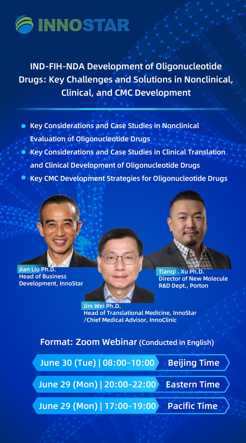
Covering three key areas—nonclinical evaluation, clinical translation, and CMC development—this webinar will examine critical decision points throughout the IND-to-FIH development process. Through practical examples and industry experience, speakers will discuss integrated strategies across pharmacology, toxicology, and CMC development, approaches to bridging preclinical and clinical development, and common pitfalls that can lead to disconnects between nonclinical and clinical programs during global regulatory submissions.
The session is designed to provide oligonucleotide development teams with a practical framework for risk assessment and decision-making, helping improve translational success from the laboratory to first-in-human studies.
Registration Link
https://forms.gle/PgKNtBTAx7jmCDjG9
Featured Speakers
Jian Liu, Ph.D.
Dr. Jian Liu has over 20 years of experience in the biopharmaceutical industry, combining scientific expertise with extensive business and leadership experience. He has led multiple projects in RNA therapeutics, siRNA-LNP delivery platforms, GLP toxicology, and IND-enabling development, supporting the translation of innovative therapies from discovery to preclinical development.
Prior to joining InnoStar, Dr. Liu served as Director of Toxicology at Nitto BioPharma, where he was responsible for nonclinical safety assessment and supported multiple IND submissions. He currently serves as Head of Business Development at InnoStar, driving global client partnerships and scientific service strategies.
Dr. Liu has published multiple peer-reviewed scientific papers and serves as Executive Vice President and Board Member of the Southern California Association for Biomedical and Pharmaceutical Advancement (SABPA), promoting collaboration between industry and academia.
Jim Wei, Ph.D.
Dr. Jim Wei is Head of Translational Medicine at InnoStar and Chief Medical Advisor at InnoClinic. He brings more than 30 years of experience in clinical pharmacology and drug development.
Dr. Wei previously served as a Senior Clinical Pharmacology Reviewer at the U.S. FDA for 10 years, where he participated in more than 100 IND, NDA, and BLA submissions and received multiple honors, including the FDA Outstanding Contribution Award and Drug Safety Award. He also spent 10 years leading early clinical development programs at Medpace and has successfully supported over 100 global early-stage drug development projects.
Dr. Wei received his Ph.D. from Idaho State University and completed postdoctoral training at the University of Washington and the National Cancer Institute (NCI/NIH). He is a Managing Partner at Milestone Biopharm, Co-founder of H&D Pharma, and a member of the American Society for Clinical Pharmacology and Therapeutics (ASCPT).
Tianqi Xu, Ph.D.
Dr. Tianqi Xu received his Ph.D. in Chemistry from The Chinese University of Hong Kong in 2016. His doctoral research focused on the synthesis of peptide analogs and the investigation of intermolecular interactions.
Over the past decade, Dr. Xu has accumulated extensive experience in peptide and oligonucleotide development, covering process development, GMP manufacturing, and CMC activities from IND-enabling studies through early clinical development. He has worked closely with both domestic and international clients and has significant expertise in the development of complex peptide therapeutics.
Dr. Xu joined Porton in May 2025 and is currently responsible for the company’s peptide and oligonucleotide business.
Address: 199 Guoshoujing Road, Pilot Free Trade Zone, Shanghai, China
Postcode: 201203
Tel: +86-21-60211999
services@innostarbio.com
86-021-xxxxxxx

Scan the QR code
Subscription to our Wechat account
Copyright © 2017-2021©Shanghai InnoStar Bio-tech Co., Ltd. All Rights Reserved. Created By VPABrand.com